【行業(yè)信息】上海藥物所徐華強課題組多巴胺受體研究成果榮登Cell封面
2021-02-20 07:50:38
2021年2月11日,上海藥物所徐華強課題組聯(lián)合國內(nèi)外多家單位于國際頂級期刊Cell以長文形式在線發(fā)表了題為Structural insights into the human D1 and D2 dopamine receptor signaling complexes的研究論文【1】,文章解析了在包括抗帕金森氏病藥物溴隱亭(bromocriptine)以及阿撲嗎啡(apomorphine)在內(nèi)的多種激動劑激活下,多巴胺受體D1R和D2R分別與下游G蛋白信號復合物的結(jié)構,并結(jié)合大量功能實驗數(shù)據(jù),揭示了D1R和D2R配體結(jié)合口袋的拓撲結(jié)構特性、潛在的受體激活機制、配體激動劑選擇性識別并激活D1R和D2R的分子機制、D1R的G蛋白偏好性激活決定因素以及D1R和D2R在G蛋白選擇性差異上的結(jié)構基礎等。
在同期Cell上,來自四川大學的邵振華團隊等以“背靠背”形式發(fā)表了Ligand recognition and allosteric regulation of DRD1-Gs signaling complexes 的研究論文【2】,報道了D1R與不同激動劑配體以及D1R與變構調(diào)節(jié)劑的結(jié)構,揭示了D1R的激動劑配體以及變構調(diào)節(jié)劑的結(jié)合特性以及潛在的變構調(diào)節(jié)機制等。
以上兩項研究成果在國際上首次分別報道了D1R的近原子分辨率結(jié)構,系統(tǒng)性地闡述了D1R的配體激動劑結(jié)合特征,為以D1R和D2R為藥物靶點的選擇性激動劑藥物的設計和開發(fā)用于治療神經(jīng)系統(tǒng)性疾病提供了重要的結(jié)構基礎和理論依據(jù)。
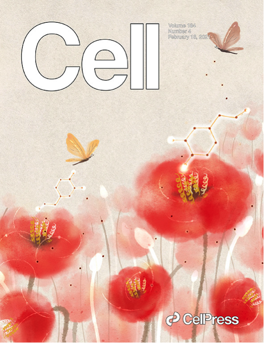
這兩項研究成果于今日在Cell以封面故事形式報道。
徐華強課題組簡介
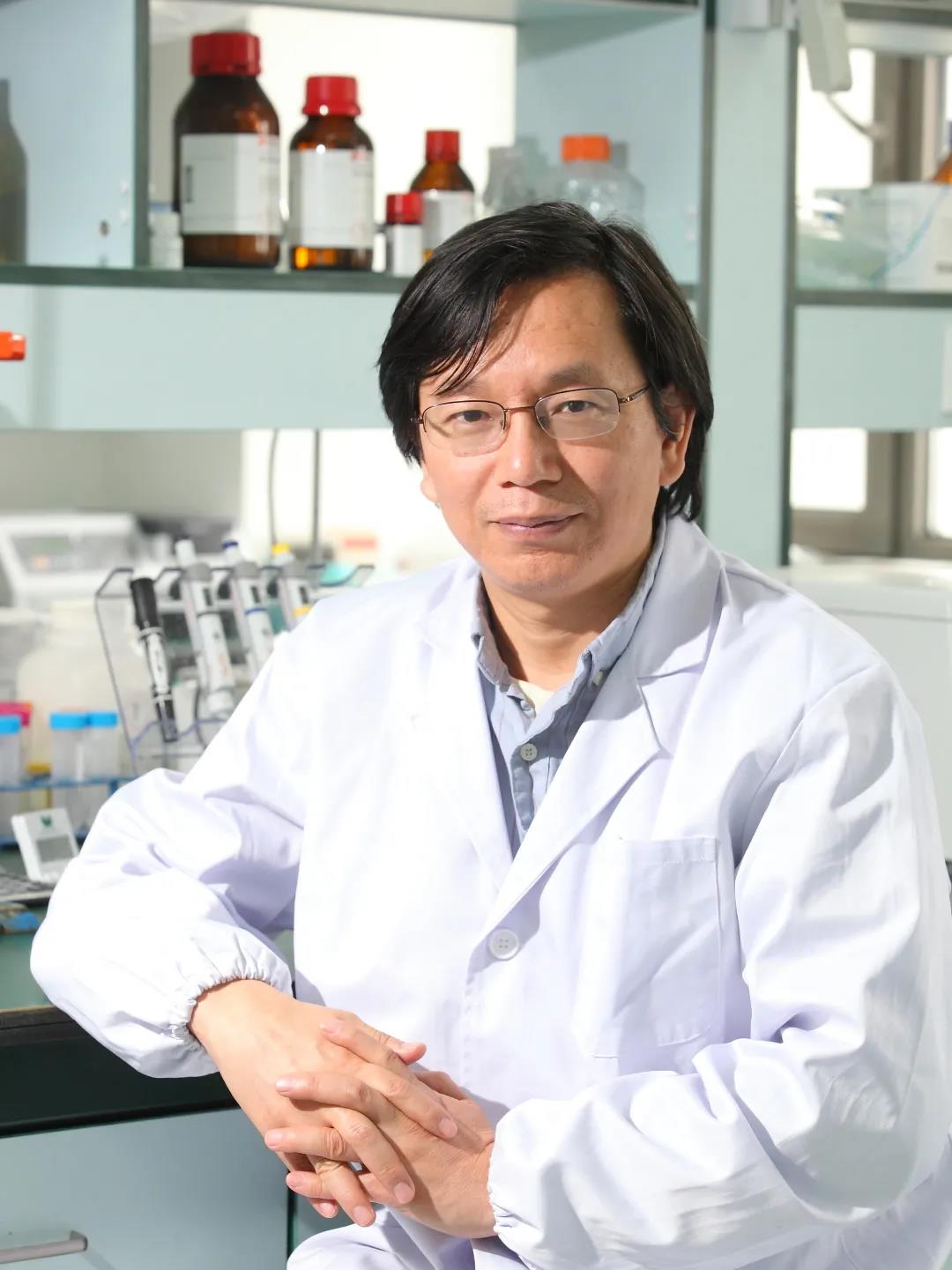
上海藥物所徐華強課題組長期致力于GPCR及其信號通路的結(jié)構和功能研究,在領域內(nèi)耕耘二十余載,取得了系列突破性成果。
近年來,課題組與國內(nèi)外科研團隊合作,解決了GPCR領域的諸多重大科學問題,創(chuàng)造了多個“首個”,包括解析國際首個GPCR與阻遏蛋白信號復合物Rhodopsin-visual arrestin的晶體結(jié)構(Yanyong. Kang, et al. 2015. Nature)【3】,首個Gi偶聯(lián)的GPCR-G蛋白信號復合物Rhodopsin-Gi(Yanyong. Kang, et al. 2018. Nature)【4】,首個非視覺阻遏蛋白(Arrestin2)與神經(jīng)降壓素受體(NTSR1)復合物(Wangchao. Yin, et al. 2019. Cell Research)【5】等。
與此同時,課題組相繼合作完成了重大疾病相關系列GPCR與G蛋白復合體的高分辨率冷凍電鏡結(jié)構及其藥理活性研究工作,包括1型人源甲狀旁腺激素受體(PTH1R)【6】、促腎上腺皮質(zhì)激素釋放激素受體(CRFRs)【7】、大麻素受體2(CB2)【8】、甲酰肽受體2(FPR2)【9 】以及黏附因子受體(GPR97)【10】 等。這些研究成果促進了人們更為深入地理解和認識GPCR信號通路,并為針對GPCR的相關藥物合理設計和開發(fā)奠定了堅實基礎,推進了基于GPCR的靶向新藥發(fā)現(xiàn)。
針對單胺類神經(jīng)遞質(zhì)受體的研究工作,徐華強課題組也開展了持續(xù)研究。早在2013年,課題組便聯(lián)合多家單位在Science上以長文形式發(fā)表了兩篇“背靠背”論文,介紹了關于五羥色胺受體家族5HT1B和5HT2B的研究成果【11】【12】。針對于多巴胺受體家族的研究,課題組還于近期在Molecular Cell上發(fā)表了D3R的研究成果【13】。
此次對于D1R和D2R的研究,是徐華強課題組在多巴胺能系統(tǒng)方向進行結(jié)構和功能系列研究的又一突出合作研究工作,進一步完善了學界對于多巴胺受體家族的認識。同時,這項工作也是在單胺類神經(jīng)遞質(zhì)受體結(jié)構藥理研究領域的又一重要進展。
參考文獻:
參考文獻:
1、Zhuang, Y. et al. Structural insights into the human D1 and D2 dopamine receptor signaling complexes. Cell, doi:10.1016/j.cell.2021.01.027 (2021).
2、Xiao, P. et al. Ligand recognition and allosteric regulation of DRD1-Gs signaling complexes. Cell, doi:10.1016/j.cell.2021.01.028 (2021).
3、Kang, Y. et al. Crystal structure of rhodopsin bound to arrestin by femtosecond X-ray laser. Nature523, 561-567, doi:10.1038/nature14656 (2015).
4、Kang, Y. et al. Cryo-EM structure of human rhodopsin bound to an inhibitory G protein. Nature558, 553-558, doi:10.1038/s41586-018-0215-y (2018).
5、Yin, W. et al. A complex structure of arrestin-2 bound to a G protein-coupled receptor. Cell research29, 971-983, doi:10.1038/s41422-019-0256-2 (2019).
6、Zhao, L. H. et al. Structure and dynamics of the active human parathyroid hormone receptor-1. Science364, 148-153, doi:10.1126/science.aav7942 (2019).
7、Ma, S. et al. Molecular Basis for Hormone Recognition and Activation of Corticotropin-Releasing Factor Receptors. Molecular cell 77, 669-680, doi:10.1016/j.molcel.2020.01.013 (2020).
8、Xing, C. et al. Cryo-EM Structure of the Human Cannabinoid Receptor CB2-Gi Signaling Complex. Cell, 180, 645-654, doi:10.1016/j.cell.2020.01.007 (2020).
9、Zhuang, Y. et al. Structure of formylpeptide receptor 2-Gi complex reveals insights into ligand recognition and signaling. Nature communications11, 885, doi:10.1038/s41467-020-14728-9 (2020).
10、Ping, Y. Q. et al. Structures of the glucocorticoid-bound adhesion receptor GPR97-Go complex. Nature589, 620-626, doi:10.1038/s41586-020-03083-w (2021).
11、Wacker, D. et al. Structural Features for Functional Selectivity at Serotonin Receptors. Science340, 615-619, doi:Doi 10.1126/Science.1232808 (2013).
12、Wang, C. et al. Structural Basis for Molecular Recognition at Serotonin Receptors. Science340, 610-614, doi:Doi 10.1126/Science.1232807 (2013).
13、Xu, P. et al. Structures of the human dopamine D3 receptor-Gi complexes. Molecular cell, doi:10.1016/j.molcel.2021.01.003 (2021).
《來源:上海藥物所》
《來源:上海藥物所》